
Chordoma - clivus
Updates to Case Attributes
Systems changed:
- Head & Neck
Updates to Study Attributes
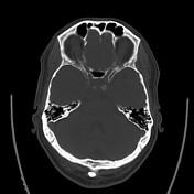
Soft tissue mass centered in the right clivus superiorly causing bony destruction. The The mass bulges into the prepontine cistern, extends into the right cavernous sinus and also extends into the posterior aspect of the sphenoid sinuses where there is rounded soft tissue density as well. Pituitary The pituitary gland and stalk look normal though there is some erosion into the posterior wall of the sella turcica present. On On transverse measurement, lesion size is approximately 2.7 x 1.3 cm. IMPRESSION: Skull base lesion centered in the clivus, causing bony destruction and likelylikely invading the right cavernous sinus. Differential Differential diagnosis includes metastatic disease, plasmacytoma, chordoma, intraosseous lymphoma. Follow Follow-up MRI assessment is indicated.
Image CT (non-contrast) ( update )
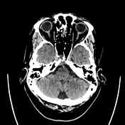
Image CT (bone window) ( update )
Updates to Study Attributes
An osteolytic solid, high T2 signal intensity, mildly heterogeneous, enhancing mass lesion is present involving the clivus as described on the recent CT. The The lesion is adjacent to the medial and posterior aspects of right internal carotid artery in the posterior cavernous sinus region, extends into the posterior aspect of the right sphenoid sinus and protrudes slightly posteriorly into the pre-pontine cistern adjacent to the anterior aspect of basilar artery. The lesion involves the posterior wall of sella but it does not involve the pituitary. IMPRESSION: An osteolytic lesion involving the clivus is present with differential diagnosis including chordoma, chondrosarcoma or metastasis. A A lymphoma or plasmacytoma could also have this appearance in the appropriate clinical setting.


 Unable to process the form. Check for errors and try again.