
Cystic hepatic metastasis
Updates to Case Attributes
This case is designed to encourage you to keep an open mind. Given the patient's history of immunosuppression, the presence of a single new new cystic lesion in the liver was definitively called an abscess on an earlier study. The patient was sent to ultrasound-guided biopsy to determine the infectious agent for targeted antibiotic therapy. The pathology report from the FNA smears and core biopsy told a different story, however:
"Metastatic, poorly-differentiated adenocarcinoma in a background of extensive necrosis and mixed inflammation."
The primary was consistent with an upper GI or pancreatic source, although this could not be seen on the available imaging.
In the differential for a new cystic lesion in in an adult liver, one should always consider both infectious and metastatic aetiologies. With multiple lesions, a metastatic aetiology is much more likely (~98%), but lack of multiple lesions is more ambiguous.
-<p>This case is designed to encourage you to keep an open mind. Given the patient's history of immunosuppression, the presence of a single new cystic lesion in the liver was definitively called an abscess on an earlier study. The patient was sent to ultrasound-guided biopsy to determine the infectious agent for targeted antibiotic therapy. The pathology report from the FNA smears and core biopsy told a different story, however:</p><p>"Metastatic, poorly-differentiated adenocarcinoma in a background of extensive necrosis and mixed inflammation."</p><p>The primary was consistent with an upper GI or pancreatic source, although this could not be seen on the available imaging.</p><p>In the differential for a new cystic lesion in an adult liver, one should always consider both infectious and metastatic aetiologies. With multiple lesions, a metastatic aetiology is much more likely (~98%), but lack of multiple lesions is more ambiguous.</p>- +<p>This case is designed to encourage you to keep an open mind. Given the patient's history of immunosuppression, the presence of a single new cystic lesion in the liver was definitively called an abscess on an earlier study. The patient was sent to ultrasound-guided biopsy to determine the infectious agent for targeted antibiotic therapy. The pathology report from the FNA smears and core biopsy told a different story, however:</p><p>"Metastatic, poorly-differentiated adenocarcinoma in a background of extensive necrosis and mixed inflammation."</p><p>The primary was consistent with an upper GI or pancreatic source, although this could not be seen on the available imaging.</p><p>In the differential for a new cystic lesion in an adult liver, one should always consider both infectious and metastatic aetiologies. With multiple lesions, a metastatic aetiology is much more likely (~98%), but lack of multiple lesions is more ambiguous.</p>
Systems changed:
- Oncology
Updates to Study Attributes
Liver CT without contrast. The study was ordered for abdominal pain. The lesion in the left hepatic lobe was new compared with a study three months earlier. There is an ill-defined hypoattenuating lesion in the lateral segment of the left hepatic lobe.
Image CT (liver window) ( update )

Image CT (non-contrast) ( update )

Updates to Study Attributes
Ultrasound correlation of the cystic liver lesion seen on CT. The lesion in the left hepatic lobe with a central hypoechoic region and a hypoechoic rim. There is only mild vascularity of the lesion on colour Doppler ultrasound.
Image Ultrasound (Transverse) ( update )
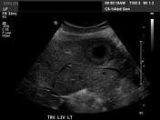
Image Ultrasound (Transverse) ( update )

Image Ultrasound (Longitudinal) ( update )

Updates to Study Attributes
Ultrasound-guided core biopsy through the solid component of the lesion.
Image Ultrasound (Transverse) ( update )


 Unable to process the form. Check for errors and try again.