
Giant liver hemangioma
Diagnosis probable
Updates to Case Attributes
Diagnostic Certainty
was set to
.
Presentation
was changed:
Race
changed from Caucasian to .
Body
was changed:
Giant haemangiomas may demonstrate similar findings to their smaller relatives, although findings are less consistent. Contrast filling may be slow and the central portions may never be demonstrated to fill in. Occasionally they may have no contrast enhancement.
This diagnosis was inferred only by imaging appearance.
-<p><a href="/articles/giant-liver-haemangioma" title="Giant liver haemangiomata">Giant haemangiomas</a> may demonstrate similar findings to their <a href="/articles/hepatic-haemangioma" title="Hepatic haemangioma">smaller relatives</a>, although findings are less consistent. Contrast filling may be slow and the central portions may never be demonstrated to fill in. Occasionally they may have no contrast enhancement.</p>- +<p><a href="/articles/giant-hepatic-venous-malformation">Giant haemangiomas</a> may demonstrate similar findings to their <a href="/articles/hepatic-haemangioma-3">smaller relatives</a>, although findings are less consistent. Contrast filling may be slow and the central portions may never be demonstrated to fill in. Occasionally they may have no contrast enhancement.</p><p>This diagnosis was inferred only by imaging appearance. </p>
Updates to Study Attributes
Caption
was added:
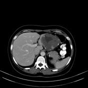
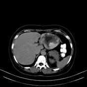
CT Abdomen
Findings
was added:
There are small hepatic cysts on the right lobe and a large hypoattenuating mass on the left lobe which shows progressive centripetal enhancement. Unfortunately this exam was performed in helical scanner and no other delayed images were obtained.
Images Changes:
Image CT (C+ arterial phase) ( update )

Perspective
was set to
Axial.
Image CT (non-contrast) ( update )

Perspective
was set to
Axial.
Image CT (C+ portal venous phase) ( update )
Perspective
was set to
Axial.
Image CT (C+ delayed) ( update )
Perspective
was set to
Axial.


 Unable to process the form. Check for errors and try again.