
Hepatic hydatid cyst
Diagnosis almost certain
Updates to Case Attributes
Diagnostic Certainty
was set to
.
Age
was set to
50.
Presentation
was changed:
Body
was changed:
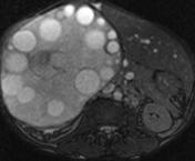
A forty seven years oldfifty year old male was referred for evaluation of large mass in liver.
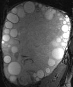
MRI showed a 24 x 17 x 18 cm multi-vesicular cyst occupying the entire right lobe of liver. Daughter cysts of varying sizes were seen predominantly in the periphery of the giant mother cyst. The collapsed membranes were seen floating within the hydatid sand.
Features are those of hydatid disease of the liver.
-<p>A forty seven years old male was referred for evaluation of  large mass in liver.</p><p>MRI showed a  24 x 17 x 18 cm multi-vesicular cyst occupying the entire right lobe of liver. Daughter cysts of varying sizes were seen predominantly in the periphery of the  giant mother cyst. The collapsed membranes were seen floating within the hydatid sand. </p><p>Features are those of <a href="/articles/hepatic-hydatid-infection" title="Hepatic hydatid cyst">hydatid disease of the liver</a>. </p>- +<p>A fifty year old male was referred for evaluation of large mass in liver.</p><p>MRI showed a 24 x 17 x 18 cm multi-vesicular cyst occupying the entire right lobe of liver. Daughter cysts of varying sizes were seen predominantly in the periphery of the giant mother cyst. The collapsed membranes were seen floating within the hydatid sand.</p><p>Features are those of <a href="/articles/hepatic-hydatid-infection">hydatid disease of the liver</a>. </p>
Updates to Study Attributes
Findings
was added:
MRI showed a 24 x 17 x 18 cm multi-vesicular cyst occupying the entire right lobe of liver. Daughter cysts of varying sizes were seen predominantly in the periphery of the giant mother cyst. The collapsed membranes were seen floating within the hydatid sand.
Images Changes:
Image MRI (T1) ( update )

Perspective
was set to
Coronal.
Single Or Stack Root
was set to
.
Image MRI (T2) ( update )

Perspective
was set to
Coronal.
Single Or Stack Root
was set to
.
Image MRI (T2) ( update )

Perspective
was set to
Coronal.
Single Or Stack Root
was set to
.
Image MRI (T2) ( update )
Perspective
was set to
Axial.
Single Or Stack Root
was set to
.
Image MRI (T2) ( update )
Perspective
was set to
Coronal.
Single Or Stack Root
was set to
.
Image MRI (T1 C+ fat sat) ( update )

Perspective
was set to
Coronal.
Single Or Stack Root
was set to
.
Specifics
changed from T2 to T1 C+ fat sat.


 Unable to process the form. Check for errors and try again.