
Lateral discoid meniscus
Diagnosis certain
Updates to Case Attributes
Age
changed from 30 to 30 years.
Updates to Study Attributes
Findings
was added:
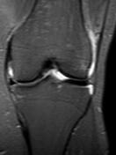
Discoid lateral meniscus.
Horizontal high T2 intrasubstance signal at the posterior horn of medial meniscus not reaching the articular surface, reflecting grade II meniscal degeneration.
Images Changes:
Image MRI (STIR) ( update )
Perspective
was set to
Coronal.
Image MRI (PD) ( update )

Stack
was set to
.
Single Or Stack Root
was set to
.
Image MRI (PD) ( update )

Stack
was set to
.
Single Or Stack Root
was set to
.
Perspective
was set to
Sagittal.


 Unable to process the form. Check for errors and try again.