
Lynch syndrome: duodenojejunal adenocarcinoma
Diagnosis almost certain
Updates to Study Attributes
Findings
was changed:
In the left upper quadrant, anterior to the renal pelvis, is a focal region of mural thickening with consistent (in a patient with known Lynch syndrome) with a DJ flexure adenocarcinoma. Note the previous colectomy.
Images Changes:
Image CT (C+ portal venous phase) ( update )

Description
was removed:
Updates to Study Attributes
Findings
was changed:
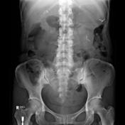
DJ flexure adenocarcinomaEvidence of prior abdominal surgery with no large bowel gas visible (total prophylactic colectomy). Apparent rounded soft tissue mass in the left upper quadrant.
Images Changes:
Image X-ray (Frontal) ( update )

Description
was removed:
Image X-ray (Frontal) ( update )
Description
was removed:
Updates to Case Attributes
Age
was set to
Young adult.
Presentation
was added:
Abdominal pain. Prior surgery.


 Unable to process the form. Check for errors and try again.