
Meningothelial hyperplasia and superficial cortical vein attenuation
Updates to Case Attributes
Meningothelial Hyperplasia (MH) is a poorly characterized entity and occasionally causes diagnostic difficulties due to its overlapping histologic features with meningioma.
MH is a reactive process characterized by a proliferation of meningothelial cells that reaches a thickness of 10ten or more cell layers with PR positivity, which is not seen in normal cells.
Brain MRI shows an remarkable accentuation of the parenchymal vessels throughout the full-thickness thickness of the cortex and whittewhite matter.
This case was presented in Eurorad, but.
Due to the authors want to share with radiopaediaMRI findings, a stereotactic biopsy was performed by the treating neurosurgical team.
-<p>Meningothelial Hyperplasia (MH) is a poorly characterized entity and occasionally causes diagnostic difficulties due to its overlapping histologic features with meningioma.</p><p>MH is a reactive process characterized by a proliferation of meningothelial cells that reaches a thickness of 10 or more cell layers with PR positivity, which is not seen in normal cells.</p><p>Brain MRI shows an remarkable accentuation of parenchymal vessels throughout the full-thickness of the cortex and whitte matter. </p><p>This case was presented in Eurorad, but the authors want to share with radiopaedia. </p>- +<p>Meningothelial Hyperplasia (MH) is a poorly characterized entity and occasionally causes diagnostic difficulties due to its overlapping histologic features with meningioma.</p><p>MH is a reactive process characterized by a proliferation of meningothelial cells that reaches a thickness of ten or more cell layers with PR positivity, which is not seen in normal cells.</p><p>Brain MRI shows remarkable accentuation of the parenchymal vessels throughout the full thickness of the cortex and white matter. </p><p>This case was presented in <em>Eurorad</em>.</p><p>Due to the MRI findings, a stereotactic biopsy was performed by the treating neurosurgical team.</p>
References changed:
- 1. Donato A, Figueroa R. Meningothelial Hyperplasia. EuroRad <a href="https://doi.org/10.1594/EURORAD/CASE.14675">doi:10.1594/EURORAD/CASE.14675</a> <span class="ref_v4"></span>
- http://www.eurorad.org/eurorad/case.php?id=14675
- Donato A, Figueroa R. Meningothelial Hyperplasia. EuroRad. Case 14675 6. href="https://http://www.eurorad.org/eurorad/case.php?id=14675">doi:10.1594/EURORAD/CASE.14675</a>
Updates to Study Attributes
MR Brain MRI demonstratedemonstrates diffuse engorgement and accentuation of parenchymlthe parenchymal vessels.
Image MRI (T1 C+) ( update )
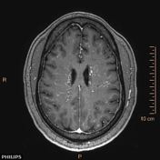
Image MRI (SWI) ( update )

Image MRI (MRA) ( update )

Image MRI (T1 C+) ( update )


 Unable to process the form. Check for errors and try again.