
Meningothelial hyperplasia and superficial cortical vein attenuation
Updates to Study Attributes
MR Brain demonstratesGadolinium-enhanced images demonstrate diffuse engorgement of multiple vascular structures extending from hemispheric cortex through subcortical white matter, deep white matter and subependymal margins on both cerebral hemispheres as well as throughout cerebellum and brainstem.
Susceptibility weighted images demonstrate remarkable accentuation of the parenchymal vessels.
There is no recognizable venous obstruction to account for vascular engorgement. There is not a dural fistula.
Image MRI (T1 C+) ( update )
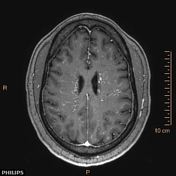
Image MRI (T1 C+) ( update )

Image MRI (SWI) ( update )

Image MRI (MRA) ( update )

Updates to Study Attributes
Cortical biopsy reports Meningothelialmeningothelial hyperplasia.
Updates to Quizquestion Attributes
Updates to Quizquestion Attributes
Updates to Case Attributes
Due to the MRI findings, a stereotactic biopsy was performed by the treating neurosurgical team.
Meningothelial hyperplasia is a poorly characterized entity and occasionally causes diagnostic difficulties due to its overlapping histologic features with meningioma. It is a reactive process characterized by a proliferation of meningothelial cells that reaches a thickness of ten or more cell layers with PR positivity, which is not seen in normal cells.
In this case, the brain MRI shows remarkable accentuation of the parenchymal vessels throughout the full thickness of the cortex and white matter.
The relationship between the vascular attenuation and the histological finding of meningothelial hyperplasia is unclear and a causal relationship has not been firmly established.
This case was presented in Eurorad.
-<p>Due to the MRI findings, a stereotactic biopsy was performed by the treating neurosurgical team.</p><p><a title="Meningothelial hyperplasia" href="/articles/meningothelial-hyperplasia">Meningothelial hyperplasia</a> is a poorly characterized entity and occasionally causes diagnostic difficulties due to its overlapping histologic features with meningioma. It is a reactive process characterized by a proliferation of meningothelial cells that reaches a thickness of ten or more cell layers with PR positivity, which is not seen in normal cells.</p><p>In this case, the brain MRI shows remarkable accentuation of the parenchymal vessels throughout the full thickness of the cortex and white matter. </p><p>The relationship between the vascular attenuation and the histological finding of meningothelial hyperplasia is unclear and a causal relationship has not been firmly established. </p><p>This case was presented in <em>Eurorad</em>.</p>- +<p>Due to the MRI findings, a stereotactic biopsy was performed by the treating neurosurgical team.</p><p><a href="/articles/meningothelial-hyperplasia">Meningothelial hyperplasia</a> is a poorly characterized entity and occasionally causes diagnostic difficulties due to its overlapping histologic features with meningioma. It is a reactive process characterized by a proliferation of meningothelial cells that reaches a thickness of ten or more cell layers with PR positivity, which is not seen in normal cells.</p><p>In this case, the brain MRI shows remarkable accentuation of the parenchymal vessels throughout the full thickness of the cortex and white matter. </p><p>The relationship between the vascular attenuation and the histological finding of meningothelial hyperplasia is unclear and a causal relationship has not been firmly established. </p><p>This case was presented in <em>Eurorad</em>.</p>


 Unable to process the form. Check for errors and try again.