
Olfactory groove meningioma - huge
Updates to Case Attributes
The patient went on to have a resection, confirming the diagnosis.
Histology
MICROSCOPIC DESCRIPTION:
The sections show a moderately cellular meningioma with attached dura. The tumour forms whorls. No sheeting arrangement is seen. The tumour cells have ovoid nuclei with no nuclear pleomorphism. Mitoses are inconspicuous. There is no necrosis. A small amount of brain parenchyma is seen and there is no evidence of brain invasion. No evidence of atypical or malignant change is identified.
FINAL DIAGNOSIS: Meningioma (WHO Grade I).
- +<p>The patient went on to have a resection, confirming the diagnosis. </p><p><strong>Histology</strong></p><p>MICROSCOPIC DESCRIPTION:</p><p>The sections show a moderately cellular meningioma with attached dura. The tumour forms whorls. No sheeting arrangement is seen. The tumour cells have ovoid nuclei with no nuclear pleomorphism. Mitoses are inconspicuous. There is no necrosis. A small amount of brain parenchyma is seen and there is no evidence of brain invasion. No evidence of atypical or malignant change is identified.</p><p>FINAL DIAGNOSIS: <a title="Meningioma - general" href="/articles/meningioma">Meningioma </a>(WHO Grade I).</p>
Updates to Study Attributes
Path pending 2215229A large hyperdense mass is present in the anterior cranial fossa, appearing to be extra-axial with a broad dural base of contact along the floor of the anterior cranial fossa and the olfactory grooves. Vivid homogeneous contrast enhancement.
Prominent hyperostosis is present as well as direct extension into the upper parts of the nasal cavity, between the middle ethmoidal air cells.
Image CT (non-contrast) ( update )

Image CT (C+ delayed) ( update )
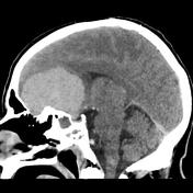
Image CT (bone window) ( update )

Image CT (bone window) ( update )
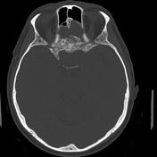
Image CT (C+ delayed) ( update )
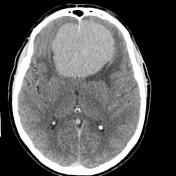
Image CT (C+ delayed) ( update )

Image CT (C+ delayed) ( update )

Image 2 CT (C+ delayed) ( update )
Image 3 CT (bone window) ( update )
Image 4 CT (C+ delayed) ( update )
Image 5 CT (C+ delayed) ( update )


 Unable to process the form. Check for errors and try again.