
Osteosarcoma in Paget disease
Updates to Case Attributes
Biopsy proven osteosarcoma.
-<p>Biopsy proven osteosarcoma</p>- +<p>Biopsy proven <a title="Osteosarcoma" href="/articles/osteosarcoma">osteosarcoma</a>. </p>
Tags changed:
- rmh
Updates to Study Attributes
CT Abdomen Pelvis
Technique: Scans were performed following oral and IV contrast (Gastrograffin and Omnipaque 350).
Findings:
Lower lumbar spine and pelvic Paget disease, left hemipelvis worse than right. In the left ilium, along the pelvic surface, enlarging enhancing soft tissue mass is again demonstrated with associated periosteal new bone formation. This has increased from 6.4 x 1.7 x 7.7 cm (07/12/2013(2 years ago) to 7.5 x 2.4 x 9.9 cm (04/02/2015(6 months ago) to currently 8 x 2.6 x 11 cm.
Conclusion:
Enlarging soft tissue mass related to Paget disease of the left hemipelvis remains concerning for sarcomatous transformation. Metastasis is felt less likely. No other potential sites of metastatic disease.
Image CT (C+ arterial phase) ( update )

Image CT (C+ arterial phase) ( update )
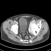
Image CT (C+ portal venous phase) ( update )

Image CT (C+ portal venous phase) ( update )
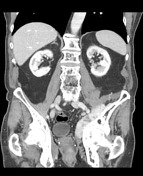
Image CT (bone window) ( update )

Image CT (bone window) ( update )

Image 4 CT (bone window) ( update )


 Unable to process the form. Check for errors and try again.