
Patellar tendinosis
Diagnosis almost certain
Updates to Case Attributes
Presentation
was changed:
Anterior knee pain -for 3 months.
? Minor, minor trauma.?
Updates to Study Attributes
Findings
was changed:
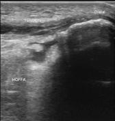
Intrasubstance fluid cleft in mid line involving proximal - mid third fibres of tendon. Fluid cleft does not involve full thickness of tendon. It does not reach to superfiicalsuperficial or deep surface of tendon.
Fibres onof medial and lateral aspect of tendon are normal.
Calcification is noted in distal third of patellar tendon fibres.
Deep infrapatellar bursal fluid is noted.
Caption
was added:
Ultrasound knee
Images Changes:
Image Ultrasound (Longitudinal) ( update )

Description
was removed:
Image Ultrasound (Transverse) ( update )

Description
was removed:
Image Ultrasound (Longitudinal) ( update )

Description
was removed:
Image Ultrasound (Longitudinal) ( update )

Description
was removed:
Image Ultrasound (Longitudinal) ( update )

Description
was removed:
Image Ultrasound (Longitudinal) ( update )
Description
was removed:
Image Ultrasound (Transverse) ( update )

Description
was removed:
Image Ultrasound (Transverse) ( update )

Description
was removed:
Image Ultrasound (Longitudinal) ( update )

Description
was removed:
Updates to Study Attributes
Caption
was added:
Knee x-ray


 Unable to process the form. Check for errors and try again.