
Perforated acute appendicitis in pregnancy (MRI)
Diagnosis certain
Disclosures
- updated 22 Aug 2022:
Nothing to disclose
Hidden edits. Some edits not affecting the appearance of this case have been suppressed.
Updates to Study Attributes
Findings
was changed:
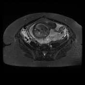
Intrauterine pregnancy confirmed but not interrogated in detail. The gravid uterus displaces the caecalcecal pole and appendix out of the right iliac fossa. An acute inflammatory mass is present adjacent to the caecalcecal pole, medially and posteriorly, with oedemaedema of the fat and a small volume of free fluid. An appendicolith isAppendicoliths are seen within the distended oedematousedematous appendix, with a small fluid collection immediately adjacent. Appearances consistent with acute appendicitis with localisedlocalized perforation and fluid collection. Incidental note is made of distension of the right ovarian vein.
Images Changes:
Image 23 MRI (STIR) ( create )
Annotation 1840
changed from ,1 arrow,1 label to fluid collection,1 arrow,1 label.
Image 23 MRI (STIR) ( create )
Annotation 1842
changed from ,1 arrow,1 label to appendicolith,1 arrow,1 label.
Image 23 MRI (STIR) ( create )
Annotation 1844
changed from ,1 arrow,1 label to stone in inflamed appendix,1 arrow,1 label.
Image 23 MRI (STIR) ( create )
Annotation 1845
changed from ,1 arrow,1 label to fluid,1 arrow,1 label.


 Unable to process the form. Check for errors and try again.