
Pituitary apoplexy
Diagnosis almost certain
Updates to Case Attributes
Diagnostic Certainty
was set to
.
Body
was changed:
Pituitary apoplexy is an acute clinical syndrome caused by either haemorrhage or infarction of thepituitary pituitary gland. Although variable, it typically comprises of headache, visual deficits, ophthalmoplegia, and altered mental status. An existing pituitary macroadenoma is usually present (60-90%) but it can occur with healthy glands in few isolated cases.
The patient had the diagnosis confirmed histologically after transsphenoidal resection.
-<p><strong>Pituitary apoplexy</strong> is an acute clinical syndrome caused by either haemorrhage or infarction of the <a href="/articles/pituitary-gland">pituitary gland</a>. Although variable, it typically comprises of headache, visual deficits, ophthalmoplegia, and altered mental status. An existing <a href="/articles/pituitary-macroadenoma-1">pituitary macroadenoma</a> is usually present (60-90%) but it can occur with healthy glands in few isolated cases.</p>- +<p><strong>Pituitary apoplexy</strong> is an acute clinical syndrome caused by either haemorrhage or infarction of the pituitary gland. Although variable, it typically comprises of headache, visual deficits, ophthalmoplegia, and altered mental status. An existing <a href="/articles/pituitary-macroadenoma-1">pituitary macroadenoma</a> is usually present (60-90%) but it can occur with healthy glands in few isolated cases.</p><p>The patient had the diagnosis confirmed histologically after transsphenoidal resection. </p>
Updates to Study Attributes
Findings
was changed:
Pituitary apoplexy
Images Changes:
Image CT (non-contrast) ( update )

Description
was removed:
Updates to Study Attributes
Findings
was changed:
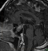
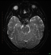
The pituitary is enlarged and heterogeneous with high T1 and low T2 signal, particularly on the right. Findings are in keeping with pituitary apoplexy.
Images Changes:
Image MRI (T1 C+) ( update )

Description
was removed:
Image MRI (T1 C+) ( update )
Description
was removed:
Image MRI (Gradient Echo) ( update )
Description
was removed:
Specifics
changed from T2 to Gradient Echo.


 Unable to process the form. Check for errors and try again.