
Pulmonary hamartoma
Diagnosis almost certain
Updates to Case Attributes
Body
was changed:
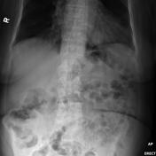
Chest x-ray demonstrates a well circumscribed opacity projecting though the cardiac silhouette.
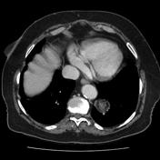
CT confirms the presence of this mass, and demonstrates that parts of it are of fat density. Features are consistent with a pulmonary hamartoma.
Follow-up has demonstrated no change in size over 5 years.
-<p>Chest x-ray demonstrates a well circumscribed opacity projecting though the cardiac silhouette.</p><p>CT confirms the presence of this mass, and demonstrates that parts of it are of fat density. Features are consistent with a <a href="/articles/pulmonary-hamartoma" title="Pulmonary Hamartoma">pulmonary hamartoma</a>. </p><p>Follow-up has demonstrated no change in size over 5 years.</p>- +<p>Follow-up has demonstrated no change in size over 5 years.</p>
Updates to Study Attributes
Findings
was added:
Well circumscribed opacity projecting though the cardiac silhouette. Calcific densities project over the right upper quadrant.
Images Changes:
Image X-ray (Frontal) ( update )

Perspective
was set to
Frontal.
Single Or Stack Root
was set to
.
Description
was removed:
Image X-ray (Frontal) ( update )
Perspective
was set to
Frontal.
Single Or Stack Root
was set to
.
Description
was removed:
Updates to Study Attributes
Findings
was added:
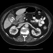
CT confirms the presence of this mass, and demonstrates that parts of it are of fat density. Features are consistent with a pulmonary hamartoma. Calcified gallstones are also noted.
Images Changes:
Image CT (C+ portal venous phase) ( update )
Perspective
was set to
Axial.
Specifics
was set to
C+ portal venous phase.
Description
was removed:
Image CT (C+ portal venous phase) ( update )
Perspective
was set to
Axial.
Specifics
was set to
C+ portal venous phase.
Description
was removed:


 Unable to process the form. Check for errors and try again.