
Pyogenic ventriculitis
Diagnosis almost certain
Updates to Case Attributes
Status
changed from draft to published (public).
Published At
was set to
.
Body
was changed:
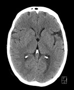
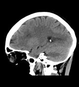
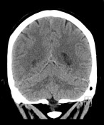
This case illustrates layering material within the lateral ventricles that almost certainly represents pus, even though there is no appreciable leptomeningeal or ependymal enhancement, which would be expected within this amount of pus.
The dilatation of the optic nerve sheaths bilaterally which can be seen in a setting of increased intracranial pressures, as can dilatation of the superior ophthalmic veins.
Blood Cultures:
POSITIVE CULTURE BOTTLE(S) 1. Streptococcus pneumoniae
Pneumococcal meningitis/ventriculitis has been suspected based on the blood culture.
-<p>This case illustrates layering material within the lateral ventricles that almost certainly represents pus, even though there is no appreciable <a title="Leptomeningeal enhancement" href="/articles/leptomeningeal-enhancement">leptomeningeal</a> or ependymal enhancement, which would be expected within this amount of pus. </p><p>The dilatation of the optic nerve sheaths bilaterally which can be seen in a setting of increased intracranial pressures, as can dilatation of the superior ophthalmic veins.</p><p> </p><p><strong>Blood Cultures:</strong></p><p>POSITIVE CULTURE BOTTLE(S) 1. <em>Streptococcus </em><em>pneumoniae</em><em> </em></p><p> </p><p>Pneumococcal meningitis/ventriculitis has been suspected based on the blood culture. </p>- +<p>This case illustrates layering material within the lateral ventricles that almost certainly represents pus, even though there is no appreciable <a href="/articles/leptomeningeal-enhancement">leptomeningeal</a> or ependymal enhancement, which would be expected within this amount of pus. </p><p>The dilatation of the optic nerve sheaths bilaterally which can be seen in a setting of increased intracranial pressures, as can dilatation of the superior ophthalmic veins.</p><p> </p><p><strong>Blood Cultures:</strong></p><p>POSITIVE CULTURE BOTTLE(S) 1. <em>Streptococcus </em><em>pneumoniae</em><em> </em></p><p> </p><p>Pneumococcal meningitis/ventriculitis has been suspected based on the blood culture. </p>
References changed:
- 1. Jorens PG, Voormolen MH, Robert D, Parizel PM. Imaging findings in pyogenic ventriculitis. Neurocritical care. 11 (3): 403-5. <a href="https://doi.org/10.1007/s12028-009-9263-3">doi:10.1007/s12028-009-9263-3</a> - <a href="https://www.ncbi.nlm.nih.gov/pubmed/19688611">Pubmed</a> <span class="ref_v4"></span>
- 2. Hong JT, Son BC, Sung JH, Kim IS, Yang SH, Lee SW, Park CK. Significance of diffusion-weighted imaging and apparent diffusion coefficient maps for the evaluation of pyogenic ventriculitis. Clinical neurology and neurosurgery. 110 (2): 137-44. <a href="https://doi.org/10.1016/j.clineuro.2007.09.019">doi:10.1016/j.clineuro.2007.09.019</a> - <a href="https://www.ncbi.nlm.nih.gov/pubmed/18023965">Pubmed</a> <span class="ref_v4"></span>
- 3. Fujikawa A, Tsuchiya K, Honya K, Nitatori T. Comparison of MRI sequences to detect ventriculitis. AJR. American journal of roentgenology. 187 (4): 1048-53. <a href="https://doi.org/10.2214/AJR.04.1923">doi:10.2214/AJR.04.1923</a> - <a href="https://www.ncbi.nlm.nih.gov/pubmed/16985156">Pubmed</a> <span class="ref_v4"></span>
Updates to Study Attributes
Modality
was set to
CT.
Caption
was added:
CT Brain (selected images)
Findings
was added:
Selected images of CT showing the appearances of the likely pus layering within the posterior horns of the lateral ventricles.
Images Changes:
Image CT (non-contrast) ( update )
Perspective
was set to
Axial.
Specifics
was set to
non-contrast.
Image CT (non-contrast) ( update )
Perspective
was set to
Sagittal.
Specifics
was set to
non-contrast.
Image CT (non-contrast) ( update )
Perspective
was set to
Coronal.
Specifics
was set to
non-contrast.
Image 1 CT (non-contrast) ( create )
Image 2 CT (non-contrast) ( create )
Image 3 CT (non-contrast) ( create )


 Unable to process the form. Check for errors and try again.