
Rickets
Diagnosis probable
Updates to Case Attributes
Body
was changed:
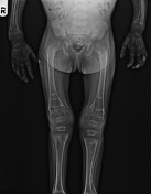
This case depicts the classical imaging findings in activerickets.
- metaphyseal fraying and cupping in knee, shoulder, elbow and wrist joints
- widening of growth plate
- generalized osteoporosis
- delayed bone age
- widening of anterior end of ribs (rachitic rosary)
- coxa vara deformity
-<p>This case depicts the classical imaging findings in active<a href="/articles/rickets"> rickets.</a></p><ul>- +<p>This case depicts the classical imaging findings in active <a href="/articles/rickets">rickets</a>.</p><ul>
Systems changed:
- Paediatrics
Updates to Study Attributes
Images Changes:
Image X-ray (Frontal) ( update )
Cropped
image
Perspective
was set to
Frontal.
Image X-ray (Frontal) ( update )

Cropped
image
Stack
was set to
.
Perspective
was set to
Frontal.


 Unable to process the form. Check for errors and try again.