
Right coronary arterial dominance with type III LAD
Diagnosis certain
Updates to Case Attributes
Title
was changed:
Age
changed from 43 to 45 years.
Status
changed from pending review to published (public).
Published At
was set to
.
Body
was changed:
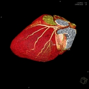
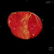
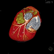
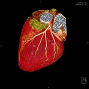
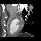
CTThis case shows an example of a right dominant coronary angiography revealscirculation with type III LAD with dominant right circulationleft anterior descending artery (LAD).
No evidence of flow-limiting stenosis noted in coronary arteries.
-<p>CT coronary angiography reveals type III LAD with dominant right circulation.</p><p>No evidence of flow-limiting stenosis noted in coronary arteries.</p>- +<p>This case shows an example of a right dominant coronary circulation with type III left anterior descending artery (LAD). </p>
Presentation
was changed:
Hypertension with hyperlipidaemia withand abnormal electrocardiogram.
Updates to Study Attributes
Findings
was changed:
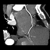
CTCT coronary angiography reveals:
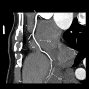
type III LAD with dominant The posterior descending artery (PDA) arises from the right circulationcoronary artery (RCA).
The left anterior descending artery (LAD) wraps around the cardiac apex into the posterior interventricular groove (type III).
No significant coronary artery stenosis identified.
Images Changes:
Image CT (C+ arterial phase) ( update )

Description
was changed:
Perspective
changed from Sagittal to Curved MPR.
Image CT (C+ arterial phase) ( update )
Description
was changed:
Perspective
changed from Sagittal to Curved MPR.
Image CT (C+ arterial phase) ( update )

Description
was changed:
Perspective
changed from Axial to Curved MPR.
Image CT (C+ arterial phase) ( update )
Description
was removed:
Specifics
changed from CT angio to C+ arterial phase.
Perspective
changed from Annotation to Volume rendered image.
Image CT (C+ arterial phase) ( update )
Description
was removed:
Perspective
changed from Annotation to Volume rendered image.
Specifics
changed from CT angio to C+ arterial phase.
Image CT (C+ arterial phase) ( update )
Description
was removed:
Perspective
changed from Annotation to Volume rendered image.
Specifics
changed from CT angio to C+ arterial phase.
Image CT (C+ arterial phase) ( update )
Description
was removed:
Perspective
changed from Annotation to Volume rendered image.
Specifics
changed from Reconstructed 3d CT angio to C+ arterial phase.
Image CT (C+ arterial phase) ( update )
Description
was changed:
Diagonal branches of the left anterior descending arteryLAD
Perspective
changed from Sagittal to Curved MPR.
Image CT (C+ arterial phase) ( update )
Perspective
changed from Sagittal to Curved MPR.
Image CT (C+ arterial phase) ( update )

Perspective
changed from Sagittal to Curved MPR.
Image CT (C+ arterial phase) ( update )

Perspective
changed from Sagittal to Curved MPR.
Image CT (C+ arterial phase) ( update )

Perspective
changed from Sagittal to Curved MPR.


 Unable to process the form. Check for errors and try again.