
Serous cystadenoma of the ovary
Updates to Case Attributes
17 year - old female with abdominal distention due to a hugeVery large, unilocular cystic mass, with no concerning morphologic features to suggest a high grade malignancy. As they are asymptomatic apart from the mass effect, it is not uncommon for benign ovarian neoplasms to be very large at presentation.
Shown to be an ovarian serous cystadenoma.
-<p>17 year - old female with abdominal distention due to a huge cystic mass. Shown to be an <a href="/articles/serous-cystadenoma" title="serous cystadenoma">ovarian serous cystadenoma</a>.</p>- +<p>Very large, unilocular cystic mass, with no concerning morphologic features to suggest a high grade malignancy. As they are asymptomatic apart from the mass effect, it is not uncommon for benign ovarian neoplasms to be very large at presentation. </p><p>Shown to be an <a href="/articles/serous-cystadenoma">ovarian serous cystadenoma</a>.</p>
Updates to Study Attributes
See belowCT confirms the presence of a large, solitary well defined abdominopelvic cystic mass lesion. There are no internal septations, solid components or papillary projections.
There is a small amount of pelvic free fluid.
Image CT (C+ portal venous phase) ( update )

Image CT (C+ portal venous phase) ( update )
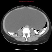
Image CT (C+ portal venous phase) ( update )

Updates to Study Attributes
See belowLarge unilocular cystic lesion, located in the midline. There are no evident solid components, papillary projections or internal septations. It is difficult to assess on ultrasound in its entirety due to the very large size. The right ovary is identified, separate to the mass. Left ovary not visualised.
Image Ultrasound ( update )

Image 1 Ultrasound ( update )


 Unable to process the form. Check for errors and try again.