
Thymoma
Updates to Study Attributes
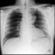
Soft tissue density projecting to the left of the mediastinum. The distal part of the arch and proximal descending aorta can be seen as separate suggesting it is located anterior to these structures.
Image X-ray (Frontal) ( update )
Updates to Study Attributes
MRI confirms the presence of an anterior mediastinal mass without convincing macroscopic invasion.
Image MRI (T1) ( update )

Image MRI (T1) ( update )

Updates to Freetext Attributes
Histology
The tumour is a thymoma of the cortical type.
The mass is firm and covered anterolaterally with a thin translucent membrane, consistent with mediastinal pleura. The cut surface is yellow-tan in colour with variably sized lobulation and focal haemorrhagic/degenerative areas in the central portion.
There is extension of the tumor into the surrounding mediastinal fat, outside of the tumor capsule, and this qualifies as an invasive thymoma. The tumor tends to push the mediastinal pleural margin and the pericardial structures. There is no evidence of pericardial involvement.


 Unable to process the form. Check for errors and try again.