
Torn discoid lateral meniscus
Diagnosis certain
Updates to Case Attributes
Diagnostic Certainty
was set to
.
Updates to Study Attributes
Findings
was changed:
The lateral meniscus is discoid iei.e. lacks the normal "bow-tie" appearance of the meniscus with a radial tear at the junction of the anterior horn and body.
Images Changes:
Image MRI (T2 fat sat) ( update )
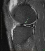
Perspective
was set to
Sagittal.
Single Or Stack Root
was set to
.
Image MRI (T2 fat sat) ( update )
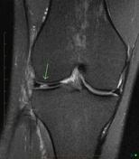
Perspective
was set to
Coronal.
Single Or Stack Root
was set to
.
Image MRI (T2 fat sat) ( update )
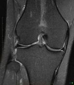
Perspective
was set to
Coronal.
Single Or Stack Root
was set to
.
Image MRI (T2 fat sat) ( update )
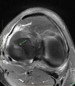
Perspective
was set to
Axial.
Single Or Stack Root
was set to
.
Specifics
was set to
T2 fat sat.


 Unable to process the form. Check for errors and try again.