
Trifurcated middle cerebral artery
Diagnosis certain
Updates to Study Attributes
Findings
was added:
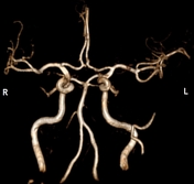
3D VRT of cerebral MR angiography shows trifurcated M2 segment left middle cerebral artery.
Images Changes:
Image Annotated image (3D VRT of circle of Willis) ( update )
Perspective
changed from Circle of Willis to 3D VRT of circle of Willis.
Updates to Study Attributes
Caption
was changed:
MR Cerebral Angiogramcerebral angiogram
Images Changes:
Image MRI (MRA) ( update )

Cropped
image
Updates to Case Attributes
Body
was changed:
The commonest pattern (around 80%) of branching of the MCA is by bifurcation of the M2 segment into superior and inferior divisions. About 10% make trifurcations and another 10% split into more than three branches1.
One One implication of such branching pattenspatterns is that the point of division is prone to aneurysms and pseudoaneurysms.
-<p>The commonest pattern (around 80%) of branching of the MCA is by bifurcation of the M2 segment into superior and inferior divisions. About 10% make trifurcations and another 10% split into more than three branches.</p><p>One implication of such branching pattens is that the point of division is prone to aneurysms and pseudoaneurysms.</p>- +<p>The commonest pattern (around 80%) of branching of the MCA is by bifurcation of the M2 segment into superior and inferior divisions. About 10% make trifurcations and another 10% split into more than three branches <sup>1</sup>. One implication of such branching patterns is that the point of division is prone to aneurysms and pseudoaneurysms.</p>
References changed:
- 1. Anderhuber F, Weiglein A, Pucher R. [Trifurcations of the Middle Cerebral Arteries]. Acta Anat (Basel). 1990;137(4):342-9. - <a href="https://www.ncbi.nlm.nih.gov/pubmed/2368589">Pubmed</a>
- Anderhuber F, Weiglein A, Pucher R. [Trifurcations of the Middle Cerebral Arteries]. Acta Anat (Basel). 1990;137(4):342-9. - <a href="https://www.ncbi.nlm.nih.gov/pubmed/2368589">Pubmed</a>


 Unable to process the form. Check for errors and try again.