


172 results found
Case
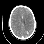
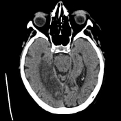
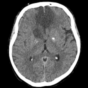
Bilateral ACA infarction due to azygos ACA embolism

Published
12 Jan 2023
93% complete
MRI
CT
Case
Acute P1 occlusion with PCA ischemia penumbra (CT perfusion)

Published
07 Nov 2019
95% complete
MRI
CT
DSA (angiography)
Case
Methamphetamine-induced intracerebral hemorrhage

Published
11 Aug 2017
89% complete
CT
DSA (angiography)
Case
Pott puffy tumor

Published
08 Sep 2019
98% complete
CT
Case
Cortical necrosis post-AMI and cardiac arrest
Published
28 Dec 2023
86% complete
CT
Case
Massive cavernous ICA aneurysm
Published
23 Oct 2023
92% complete
CT
Case
Insula and operculum (annotated MRI)

Published
03 Apr 2019
22% complete
Annotated image
Case
Cerebral abscess with ventriculitis

Published
17 Jun 2020
92% complete
MRI
CT
Case
Brain lobes - annotated MRI

Published
14 Jul 2018
15% complete
MRI
Case
CT head axial - labeling questions

Published
16 Aug 2018
40% complete
CT
Annotated image
Case
Vein of Trolard thrombosis with venous infarction (CT perfusion)

Published
20 Jul 2020
94% complete
CT
MRI
Case
Ischemic stroke - dense posterior cerebral artery (PCA) sign

Published
08 Sep 2019
95% complete
CT
Case
Dural venous sinuses (Gray's illustrations)

Published
20 Dec 2021
32% complete
Diagram
Case
Cerebral abscess secondary to mastoiditis
Published
18 Oct 2023
92% complete
MRI
Case
Möbius syndrome

Published
08 Sep 2019
80% complete
MRI
Case
Type II proatlantal artery

Published
05 Sep 2019
95% complete
CT
Case
Venous vascular territories of the medial cerebral cortex (illustration)

Published
20 Jul 2020
44% complete
Diagram
Case
Craniectomy bone plate in abdominal wall

Published
29 Nov 2019
83% complete
CT
Case
Orbital apex (diagram)

Published
04 Apr 2017
38% complete
Diagram
Case
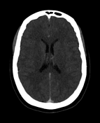
Diffuse hypoxic ischemic brain injury

Published
13 Apr 2020
95% complete
CT
Case
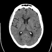
Normal head (CT perfusion)

Published
19 Nov 2019
85% complete
CT
Case
Normal head CT venogram
Published
22 Dec 2022
83% complete
CT
Case
PCA infarction
Published
15 Apr 2023
92% complete
CT
Case
Pericallosal ACA infarction
Published
15 Apr 2023
89% complete
CT
MRI
Case
Cortical vein thrombosis with infarct and hemorrhage

Published
23 Jul 2020
95% complete
MRI
CT
Annotated image
Case
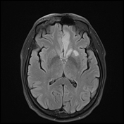
Susac syndrome

Published
04 Dec 2019
77% complete
MRI
Case
Pacemaker and brainstem stimulators (chest x-ray)

Published
28 Jun 2019
85% complete
X-ray
Case
Anatomical planes (creative commons illustration)
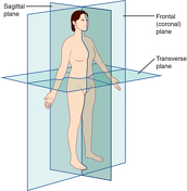
Published
20 Mar 2018
32% complete
Diagram
Case
Anatomical relations (creative commons illustration)

Published
20 Mar 2018
32% complete
Diagram
Case
Venous infarct due to superior sagittal sinus and superior cortical vein thrombosis
Published
12 Jan 2023
95% complete
CT
MRI
Case
Neurofibromatosis skin lesions on CXR

Published
10 Jun 2020
88% complete
X-ray
Case
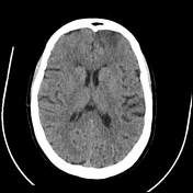
Bilateral ACA infarction
Published
19 Jan 2023
92% complete
CT
Case
ACA orbitofrontal infarct post DSA
Published
11 Jan 2023
89% complete
CT
DSA (angiography)
Case
Acquired hepatocerebral degeneration

Published
27 Jan 2021
83% complete
MRI
Case
Gyrus rectus, cingulate gyrus and caudate head infarction post ACA aneurysm coiling
Published
15 Feb 2023
95% complete
MRI
Case
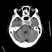
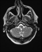
Lateral medullary infarct
Published
15 Feb 2023
89% complete
MRI
Case
Posterior limb internal capsule infarct after MCA aneurysm coiling
Published
15 Feb 2023
90% complete
CT
Case
Hippocampal and thalamic infarction from vertebral artery dissecting aneursym
Published
15 Feb 2023
84% complete
CT
MRI
Case
Hummingbird (photo)

Published
23 Apr 2020
32% complete
Photo
Case
Cutaneous spinal nerves of the lower limb (Gray's illustrations)

Published
05 Jan 2022
29% complete
Diagram
Case
Cutaneous spinal nerves of the upper limb (Gray's illustrations)

Published
05 Jan 2022
29% complete
Diagram
Case
Brachial plexus (Gray's illustrations)

Published
05 Jan 2022
32% complete
Diagram
Case
Dermatomes (Gray's illustrations)
Published
05 Jan 2022
35% complete
Diagram
Case
Spinal nerve roots (Gray's illustrations)

Published
05 Jan 2022
32% complete
Diagram
Case
Cervical plexus (Gray's illustrations)

Published
05 Jan 2022
32% complete
Diagram
Case
Basal ganglia (Gray's illustrations)
Published
27 Dec 2021
32% complete
Diagram
Case
Corona radiata (Gray's illustration)

Published
27 Dec 2021
32% complete
Diagram
Case
Internal capsule fibers (Gray's illustration)

Published
27 Dec 2021
32% complete
Diagram
Case
Tela choroidea and choroid plexus of lateral ventricles (Gray's illustration)

Published
27 Dec 2021
44% complete
Diagram
Case
Hippocampus (Gray's illustration)
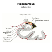
Published
27 Dec 2021
32% complete
Diagram
Case
Fornix (Gray's illustration)

Published
27 Dec 2021
32% complete
Diagram
Case
Internal features of the lateral ventricles (Gray's illustrations)

Published
27 Dec 2021
32% complete
Diagram
Case
Corpus striatum (Gray's illustration)

Published
27 Dec 2021
35% complete
Diagram
Case
Cavernous sinus (Gray's illustration)

Published
08 Dec 2021
32% complete
Diagram
Case
Internal cerebral veins (Gray's illustration)

Published
07 Dec 2021
32% complete
Diagram
Case
CT angiogram head sagittal - labeling questions

Published
30 Nov 2021
40% complete
Annotated image
CT
Case
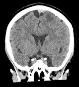
CT angiogram head coronal - labeling questions

Published
29 Nov 2021
40% complete
CT
Annotated image
Case
MRI pituitary gland coronal T1 post contrast - labeling questions

Published
17 Nov 2021
40% complete
Annotated image
MRI
Case
MRI pituitary gland sagittal T1 post contrast - labeling questions

Published
16 Nov 2021
40% complete
MRI
Annotated image
Case
MRI head axial T2 - labeling questions

Published
02 Nov 2021
40% complete
Annotated image
MRI
Case
MRI head sagittal T1 - labeling questions

Published
28 Oct 2021
40% complete
Annotated image
MRI
Case
Accessory PCA arising from the terminal ICA

Published
08 Nov 2021
79% complete
MRI
Case
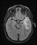
Superior ophthalmic vein thrombosis

Published
31 Aug 2021
92% complete
CT
Case
Subdural hemorrhage on CT perfusion

Published
26 Aug 2021
39% complete
CT
Case
Toxic encephalopathy

Published
19 May 2021
65% complete
MRI
Case
Subependymal giant cell astrocytoma (SEGA)

Published
03 May 2021
74% complete
MRI
Case
Cerebral fat embolism

Published
02 Jan 2021
81% complete
CT
X-ray
MRI
Case
CT angiogram head axial - labeling questions

Published
24 Nov 2020
40% complete
CT
Annotated image
Case
Toxic encephalopathy

Published
19 May 2021
59% complete
MRI
Case
Brainstem arterial territories (diagrams)

Published
07 Oct 2020
44% complete
Diagram
Case
Accessory articulation of cervical transverse processes

Published
03 Oct 2020
74% complete
Annotated image
CT
Case
Mandibular division of the trigeminal nerve and submandibular and otic ganglia (Gray's illustration)
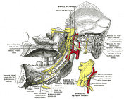
Published
20 Sep 2020
35% complete
Diagram
Case
Mandibular division of the trigeminal nerve (Gray's illustration)
Published
20 Sep 2020
35% complete
Diagram
Case
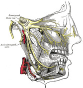
Maxillary division of the trigeminal nerve (Gray's illustration)

Published
20 Sep 2020
35% complete
Diagram
Case
Maxillary and mandibular divisions of the trigeminal nerve (Gray's illustration)
Published
20 Sep 2020
35% complete
Diagram
Case
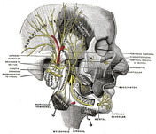
Nerves of the face, scalp and neck (Gray's illustration)
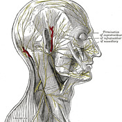
Published
20 Sep 2020
35% complete
Diagram
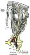
Case
Anatomy of the genicular ganglion (Gray's illustration)

Published
19 Sep 2020
35% complete
Diagram
Case
Nerves of the orbit (Gray's illustration)

Published
19 Sep 2020
35% complete
Diagram
Case
Anatomy of the ophthalmic division of the trigeminal nerve (Gray's illustration)
Published
19 Sep 2020
35% complete
Diagram
Case
Anatomy of the oculomotor nerve (Gray's illustration)

Published
19 Sep 2020
35% complete
Diagram
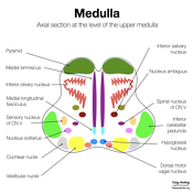
Case
Cranial nerve nuclei (axial diagrams)

Published
18 Sep 2020
25% complete
Diagram
Case
Pontine anatomy - CN V (diagram)

Published
18 Sep 2020
32% complete
Diagram
Case
Brainstem cross-sectional anatomy (diagrams)

Published
08 Sep 2020
29% complete
Diagram
Case
Lower pons anatomy - CN VII (diagram)

Published
18 Aug 2020
22% complete
Diagram
Case
Lower pons anatomy - CN VI (diagram)

Published
18 Aug 2020
22% complete
Diagram
Case
Olfactory nerve (Gray's illustration)

Published
17 Sep 2020
35% complete
Diagram
Case
Optic nerve and chiasm (Gray's illustration)

Published
17 Sep 2020
35% complete
Diagram
Case
Upper medulla anatomy - CN XII (diagram)

Published
17 Aug 2020
32% complete
Diagram
Case
Upper medulla anatomy - CN XI (diagram)

Published
17 Aug 2020
32% complete
Diagram
Case
Upper medulla anatomy - CN VIII (diagram)

Published
01 Sep 2020
32% complete
Diagram
Case
Upper medulla anatomy - CN IX (diagram)

Published
01 Sep 2020
32% complete
Diagram
Case
Upper medulla anatomy - CN X (diagram)
Published
02 Sep 2020
22% complete
Diagram
Case
Cranial meninges and falx (Gray's illustration)

Published
14 Sep 2020
35% complete
Diagram
ADVERTISEMENT: Supporters see fewer/no ads


 Unable to process the form. Check for errors and try again.