


171 results found
Case
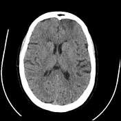
ACA orbitofrontal infarct post DSA
Published
11 Jan 2023
89% complete
CT
DSA (angiography)
Case
Accessory articulation of cervical transverse processes

Published
03 Oct 2020
74% complete
Annotated image
CT
Case
Accessory PCA arising from the terminal ICA

Published
08 Nov 2021
79% complete
MRI
Case
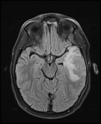
Acquired hepatocerebral degeneration

Published
27 Jan 2021
83% complete
MRI
Case
Acute A3 occlusion with ACA ischemic penumbra (CT perfusion)

Published
05 Nov 2019
94% complete
CT
Case
Acute aneurysmal SAH complicated by vasospasm

Published
30 Aug 2022
86% complete
CT
Case
Acute ICA ischemic penumbra due to high-grade CCA stenosis (CT perfusion)

Published
05 Nov 2019
94% complete
CT
Case
Acute M1 occlusion with ischemic penumbra (CT perfusion)

Published
29 Oct 2019
94% complete
DSA (angiography)
CT
Case
Acute P1 occlusion with PCA ischemia penumbra (CT perfusion)

Published
07 Nov 2019
95% complete
MRI
CT
DSA (angiography)
Case
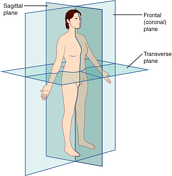
Anatomical planes (creative commons illustration)
Published
20 Mar 2018
32% complete
Diagram
Case
Anatomical relations (creative commons illustration)

Published
20 Mar 2018
32% complete
Diagram
Case
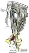
Anatomy of the genicular ganglion (Gray's illustration)

Published
19 Sep 2020
35% complete
Diagram
Case
Anatomy of the lateral ventricles (Gray's illustration)

Published
03 Sep 2020
32% complete
Diagram
Case
Anatomy of the oculomotor nerve (Gray's illustration)

Published
19 Sep 2020
35% complete
Diagram
Case
Anatomy of the ophthalmic division of the trigeminal nerve (Gray's illustration)
Published
19 Sep 2020
35% complete
Diagram
Case
Aplastic A1 on MRA

Published
24 Aug 2018
61% complete
MRI
Case
Ascending spinal tracts (Gray's illustration)

Published
08 Sep 2020
35% complete
Diagram
Case
Autonomic ganglia of the head and neck (Gray's illustrations)

Published
13 Oct 2022
35% complete
Diagram
Case
Autonomic nervous system (Gray's illustration)

Published
12 Oct 2022
35% complete
Diagram
Case
Basal ganglia chronic hypoxic ischemic disease

Published
13 Apr 2020
95% complete
CT
MRI
Case
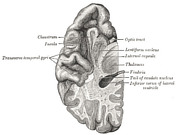
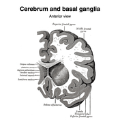
Basal ganglia (Gray's illustration)
Published
14 Sep 2020
35% complete
Diagram
Case
Basal ganglia (Gray's illustrations)
Published
27 Dec 2021
32% complete
Diagram
Case
Bilateral ACA infarction
Published
19 Jan 2023
92% complete
CT
Case
Bilateral ACA infarction due to azygos ACA embolism

Published
12 Jan 2023
93% complete
MRI
CT
Case
Bilateral cerebellar tonsil infarction

Published
11 Oct 2022
95% complete
CT
MRI
Case
Bilateral choroid plexus xanthogranulomata

Published
06 Jan 2020
74% complete
CT
Case
Brachial plexus (Gray's illustrations)

Published
05 Jan 2022
32% complete
Diagram
Case
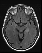
Brain lobes - annotated MRI

Published
14 Jul 2018
15% complete
MRI
Case
Brain perfusion - time attenuation curves

Published
13 Aug 2019
29% complete
Diagram
Case
Brainstem arterial territories (diagrams)

Published
07 Oct 2020
44% complete
Diagram
Case
Brainstem cross-sectional anatomy (diagrams)

Published
08 Sep 2020
29% complete
Diagram
Case
Brainstem tracts (Gray's illustrations)

Published
06 Sep 2020
32% complete
Diagram
Case
Brain venous vascular territories (diagram)

Published
16 Jul 2020
44% complete
Diagram
Case
Buffalo pneumothorax - post-operative

Published
07 Feb 2019
94% complete
X-ray
Case
Calcified chronic subdural hematoma
Published
15 May 2020
92% complete
CT
Case
Cauda equina (Gray's illustration)

Published
07 Sep 2020
35% complete
Diagram
Case
Cavernous sinus (Gray's illustration)

Published
08 Dec 2021
32% complete
Diagram
Case
Central vein and iron rim sign

Published
06 Aug 2020
92% complete
MRI
Case
Cerebellar peduncles (Gray's illustration)

Published
03 Sep 2020
35% complete
Diagram
Case
Cerebellar peduncles (Gray's illustration)

Published
03 Sep 2020
35% complete
Diagram
Case
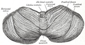
Cerebellum inferior surface (Gray's illustration)

Published
03 Sep 2020
35% complete
Diagram
Case
Cerebellum superior surface (Gray's illustration)
Published
03 Sep 2020
32% complete
Diagram
Case
Cerebral abscess secondary to mastoiditis
Published
18 Oct 2023
92% complete
MRI
Case
Cerebral abscess with ventriculitis

Published
17 Jun 2020
92% complete
MRI
CT
Case
Cerebral fat embolism

Published
02 Jan 2021
81% complete
CT
X-ray
MRI
Case
Cervical plexus (Gray's illustrations)

Published
05 Jan 2022
32% complete
Diagram
Case
Circle of Willis (Gray's illustration)

Published
06 Sep 2020
32% complete
Diagram
Case
CNS cryptococcosis

Published
08 Sep 2019
84% complete
CT
MRI
Case
Codman Hakim programmable VP shunt

Published
12 Apr 2017
88% complete
X-ray
Case
Colliculi connections (Gray's illustration)

Published
03 Sep 2020
35% complete
Diagram
Case
Corona radiata (Gray's illustration)

Published
27 Dec 2021
32% complete
Diagram
Case
Corpus striatum (Gray's illustration)

Published
27 Dec 2021
35% complete
Diagram
Case
Cortical necrosis post-AMI and cardiac arrest
Published
28 Dec 2023
86% complete
CT
Case
Cortical vein thrombosis with infarct and hemorrhage

Published
23 Jul 2020
95% complete
MRI
CT
Annotated image
Case
Cranial meninges and falx (Gray's illustration)

Published
14 Sep 2020
35% complete
Diagram
Case
Cranial nerve nuclei (axial diagrams)

Published
18 Sep 2020
25% complete
Diagram
Case
Cranial nerves (Gray's illustration)

Published
08 Sep 2020
32% complete
Diagram
Case
Cranial nerves in the posterior fossa (Gray's illustration)

Published
06 Sep 2020
35% complete
Diagram
Case
Craniectomy bone plate in abdominal wall

Published
29 Nov 2019
83% complete
CT
Case
Crossed cerebellar diaschisis

Published
23 Dec 2018
77% complete
CT
Case
CT angiogram head axial - labeling questions

Published
24 Nov 2020
40% complete
CT
Annotated image
Case
CT angiogram head coronal - labeling questions

Published
29 Nov 2021
40% complete
CT
Annotated image
Case
CT angiogram head sagittal - labeling questions

Published
30 Nov 2021
40% complete
Annotated image
CT
Case
CT head axial - labeling questions

Published
16 Aug 2018
40% complete
CT
Annotated image
Case
CT head axial - labeling quiz

Published
23 Aug 2018
15% complete
CT
Case
CT head coronal - labeling questions

Published
26 Aug 2018
40% complete
Annotated image
CT
Case
CT head sagittal - labeling questions

Published
23 Aug 2018
40% complete
CT
Annotated image
Case
Cutaneous spinal nerves of the lower limb (Gray's illustrations)

Published
05 Jan 2022
29% complete
Diagram
Case
Cutaneous spinal nerves of the upper limb (Gray's illustrations)

Published
05 Jan 2022
29% complete
Diagram
Case
Decussation of fibers in the brainstem (Gray's illustration)

Published
03 Sep 2020
35% complete
Diagram
Case
Dentate nucleus (Gray's illustration)

Published
03 Sep 2020
32% complete
Diagram
Case
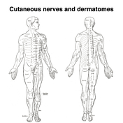
Dermatomes (Gray's illustrations)
Published
05 Jan 2022
35% complete
Diagram
Case
Descending spinal tracts (Gray's illustration)

Published
08 Sep 2020
35% complete
Diagram
Case
Diffuse hypoxic ischemic brain injury

Published
13 Apr 2020
95% complete
CT
Case
Dural venous sinuses (Gray's illustrations)

Published
20 Dec 2021
32% complete
Diagram
Case
Dural venous sinus thrombosis

Published
31 May 2018
80% complete
CT
Case
Fornix (Gray's illustration)

Published
27 Dec 2021
32% complete
Diagram
Case
Gyrus rectus, cingulate gyrus and caudate head infarction post ACA aneurysm coiling
Published
15 Feb 2023
95% complete
MRI
Case
Hippocampal and thalamic infarction from vertebral artery dissecting aneursym
Published
15 Feb 2023
84% complete
CT
MRI
Case
Hippocampus (Gray's illustration)
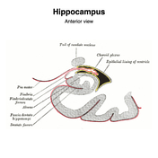
Published
27 Dec 2021
32% complete
Diagram
Case
Hummingbird (photo)

Published
23 Apr 2020
32% complete
Photo
Case
Ice cream cone (photo)

Published
30 Apr 2020
35% complete
Photo
Case
Insula and operculum (annotated MRI)

Published
03 Apr 2019
22% complete
Annotated image
Case
Insular cortex (Gray's illustration)

Published
14 Sep 2020
32% complete
Diagram
Case
Interconnection of cranial nerve nuclei (Gray's illustration)

Published
03 Sep 2020
35% complete
Diagram
Case
Interhemispheric fissure

Published
13 May 2018
37% complete
CT
MRI
Case
Internal capsule fibers (Gray's illustration)

Published
27 Dec 2021
32% complete
Diagram
Case
Internal cerebral veins (Gray's illustration)

Published
07 Dec 2021
32% complete
Diagram
Case
Internal features of the lateral ventricles (Gray's illustrations)

Published
27 Dec 2021
32% complete
Diagram
Case
Intracardiac VP shunt migration

Published
23 May 2019
95% complete
CT
DSA (angiography)
Case
Ischemic penumbra and core infarct in CT perfusion

Published
02 Dec 2019
25% complete
Annotated image
Case
Ischemic stroke - dense posterior cerebral artery (PCA) sign

Published
08 Sep 2019
95% complete
CT
Case
Large orbital extraconal hematoma causing proptosis

Published
08 Jul 2020
92% complete
CT
ADVERTISEMENT: Supporters see fewer/no ads


 Unable to process the form. Check for errors and try again.