


171 results found
Case
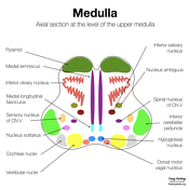
Mid-level medulla anatomy (Gray's illustration)

Published
03 Sep 2020
32% complete
Diagram
Case
Decussation of fibers in the brainstem (Gray's illustration)

Published
03 Sep 2020
35% complete
Diagram
Case
Upper medulla anatomy - CN X (diagram)
Published
02 Sep 2020
22% complete
Diagram
Case
Upper medulla anatomy - CN IX (diagram)

Published
01 Sep 2020
32% complete
Diagram
Case
Upper medulla anatomy - CN VIII (diagram)

Published
01 Sep 2020
32% complete
Diagram
Case
Lower pons anatomy - CN VII (diagram)

Published
18 Aug 2020
22% complete
Diagram
Case
Lower pons anatomy - CN VI (diagram)

Published
18 Aug 2020
22% complete
Diagram
Case
Upper medulla anatomy - CN XII (diagram)

Published
17 Aug 2020
32% complete
Diagram
Case
Upper medulla anatomy - CN XI (diagram)

Published
17 Aug 2020
32% complete
Diagram
Case
Central vein and iron rim sign

Published
06 Aug 2020
92% complete
MRI
Case
Cortical vein thrombosis with infarct and hemorrhage

Published
23 Jul 2020
95% complete
MRI
CT
Annotated image
Case
Normal brain PET

Published
23 Jul 2020
72% complete
Nuclear medicine
Case
Venous vascular territories of the lateral cerebral cortex (illustration)

Published
20 Jul 2020
44% complete
Diagram
Case
Venous vascular territories of the medial cerebral cortex (illustration)

Published
20 Jul 2020
44% complete
Diagram
Case
Vein of Trolard thrombosis with venous infarction (CT perfusion)

Published
20 Jul 2020
94% complete
CT
MRI
Case
Brain venous vascular territories (diagram)

Published
16 Jul 2020
44% complete
Diagram
Case
Large orbital extraconal hematoma causing proptosis

Published
08 Jul 2020
92% complete
CT
Case
Cerebral abscess with ventriculitis

Published
17 Jun 2020
92% complete
MRI
CT
Case
Neurofibromatosis skin lesions on CXR

Published
10 Jun 2020
88% complete
X-ray
Case
Calcified chronic subdural hematoma
Published
15 May 2020
92% complete
CT
Case
Ice cream cone (photo)

Published
30 Apr 2020
35% complete
Photo
Case
Raccoon eyes (photo)

Published
28 Apr 2020
35% complete
Photo
Case
Viking helmet (photo)

Published
23 Apr 2020
35% complete
Photo
Case
Raccoon (photo)

Published
23 Apr 2020
32% complete
Photo
Case
Leopard (photo)

Published
23 Apr 2020
35% complete
Photo
Case
Hummingbird (photo)

Published
23 Apr 2020
32% complete
Photo
Case
Moose head (photo)

Published
23 Apr 2020
41% complete
Photo
Case
Basal ganglia chronic hypoxic ischemic disease

Published
13 Apr 2020
95% complete
CT
MRI
Case
Diffuse hypoxic ischemic brain injury

Published
13 Apr 2020
95% complete
CT
Case
Spinal hemangioblastoma

Published
05 Feb 2020
89% complete
CT
MRI
Fluoroscopy
Case
Wireless intracranial pressure monitor

Published
18 Jan 2020
75% complete
X-ray
Case
Phantom head CT

Published
13 Jan 2020
21% complete
CT
Photo
Case
Bilateral choroid plexus xanthogranulomata

Published
06 Jan 2020
74% complete
CT
Case
Mastoid foramina

Published
12 Dec 2019
53% complete
CT
Case
Susac syndrome

Published
04 Dec 2019
77% complete
MRI
Case
Ischemic penumbra and core infarct in CT perfusion

Published
02 Dec 2019
25% complete
Annotated image
Case
Craniectomy bone plate in abdominal wall

Published
29 Nov 2019
83% complete
CT
Case
Malpositioned external femoral compression device with large retroperitoneal hemorrhage following femoral puncture

Published
28 Nov 2019
89% complete
CT
Case
Salted pretzel sign (CT head)

Published
22 Nov 2019
74% complete
CT
Case
Normal head (CT perfusion)

Published
19 Nov 2019
85% complete
CT
Case
Acute P1 occlusion with PCA ischemic penumbra (CT perfusion)

Published
07 Nov 2019
95% complete
CT
DSA (angiography)
MRI
Case
Acute ICA ischemic penumbra due to high-grade CCA stenosis (CT perfusion)

Published
05 Nov 2019
94% complete
CT
Case
Acute A3 occlusion with ACA ischemic penumbra (CT perfusion)

Published
05 Nov 2019
94% complete
CT
Case
Acute M1 occlusion with ischemic penumbra (CT perfusion)

Published
29 Oct 2019
94% complete
DSA (angiography)
CT
Case
Wernicke encephalopathy

Published
29 Oct 2019
71% complete
MRI
Case
Tectal plate cavernoma

Published
10 Sep 2019
82% complete
CT
MRI
Case
Multiple cerebral cavernous malformations
Published
09 Sep 2019
98% complete
MRI
CT
Case
CNS cryptococcosis

Published
08 Sep 2019
84% complete
CT
MRI
Case
Ischemic stroke - dense posterior cerebral artery (PCA) sign

Published
08 Sep 2019
95% complete
CT
Case
Pott puffy tumor

Published
08 Sep 2019
98% complete
CT
Case
Möbius syndrome

Published
08 Sep 2019
80% complete
MRI
Case
Type II proatlantal artery

Published
05 Sep 2019
95% complete
CT
Case
Brain perfusion - time attenuation curves

Published
13 Aug 2019
29% complete
Diagram
Case
Pacemaker and brainstem stimulators (chest x-ray)

Published
28 Jun 2019
85% complete
X-ray
Case
Intracardiac VP shunt migration

Published
23 May 2019
95% complete
CT
DSA (angiography)
Case
Insula and operculum (annotated MRI)

Published
03 Apr 2019
22% complete
Annotated image
Case
Right mesial temporal lobe jet hematoma from ruptured PCom artery aneurysm

Published
26 Feb 2019
95% complete
CT
Case
Buffalo pneumothorax - post-operative

Published
07 Feb 2019
94% complete
X-ray
Case
Crossed cerebellar diaschisis

Published
23 Dec 2018
77% complete
CT
Case
CT head coronal - labeling questions

Published
26 Aug 2018
40% complete
Annotated image
CT
Case
Aplastic A1 on MRA

Published
24 Aug 2018
61% complete
MRI
Case
CT head axial - labeling quiz

Published
23 Aug 2018
15% complete
CT
Case
CT head sagittal - labeling questions

Published
23 Aug 2018
40% complete
CT
Annotated image
Case
CT head axial - labeling questions

Published
16 Aug 2018
40% complete
CT
Annotated image
Case
Brain lobes - annotated MRI

Published
14 Jul 2018
15% complete
MRI
Case
Dural venous sinus thrombosis

Published
31 May 2018
80% complete
CT
Case
Progressive multifocal leukoencephalopathy immune reconstitution inflammatory syndrome

Published
24 May 2018
92% complete
MRI
Case
Interhemispheric fissure

Published
13 May 2018
37% complete
CT
MRI
Case
Anatomical relations (creative commons illustration)

Published
20 Mar 2018
32% complete
Diagram
Case
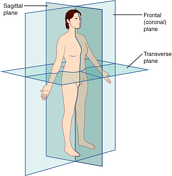
Anatomical planes (creative commons illustration)
Published
20 Mar 2018
32% complete
Diagram
Case
Vagal nerve stimulator

Published
16 Feb 2018
82% complete
X-ray
Case
Methamphetamine-induced intracerebral hemorrhage

Published
11 Aug 2017
89% complete
CT
DSA (angiography)
Case
Pulmonary alveolar microlithiasis

Published
31 May 2017
91% complete
X-ray
Case
Sophysa Polaris SPV programmable VP shunt
Published
14 Apr 2017
88% complete
X-ray
Case
Codman Hakim programmable VP shunt

Published
12 Apr 2017
88% complete
X-ray
Case
Orbital apex (diagram)

Published
04 Apr 2017
38% complete
Diagram
Case
Medtronic Strata programmable VP shunt

Published
12 Aug 2016
88% complete
X-ray
Case
Sarcoidosis

Published
17 Jul 2015
91% complete
X-ray
ADVERTISEMENT: Supporters see fewer/no ads


 Unable to process the form. Check for errors and try again.