


24 results found
Case
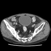
Right hemipelvis metastases simulating Paget disease
Published
17 Aug 2018
89% complete
CT
Annotated image
Case
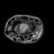
Tibialis posterior tendon rupture and sinus tarsi synovitis

Published
17 Aug 2018
77% complete
Annotated image
MRI
Case
Knee lipohemarthrosis due to tibial plateau fracture

Published
01 May 2015
89% complete
Annotated image
X-ray
CT
Case
Stener lesion

Published
21 Jan 2015
92% complete
Annotated image
MRI
Case
Osteochondral fracture of the talar dome

Published
18 Jan 2015
88% complete
Annotated image
X-ray
Diagram
Case
Pelvic fractures (Malgaigne fracture)

Published
18 Oct 2014
92% complete
Annotated image
X-ray
CT
Case
Paget disease: with blade of grass sign

Published
03 Aug 2014
91% complete
Annotated image
X-ray
CT
MRI
Nuclear medicine
Case
Peroneus longus tendon rupture associated with an os peroneum

Published
26 Jul 2014
92% complete
Annotated image
X-ray
MRI
Case
Giant cell tumor of tendon sheath

Published
16 Jul 2014
72% complete
Annotated image
Ultrasound
X-ray
Case
Ischial tuberosity avulsion

Published
11 Jul 2014
83% complete
X-ray
Annotated image
Case
Ulcerative colitis with lead pipe appearance and arthropathy

Published
31 Mar 2014
94% complete
X-ray
Annotated image
Case
Internal impingement of the shoulder

Published
17 Aug 2013
92% complete
MRI
Annotated image
Case
Supra-acetabular fossa

Published
09 Aug 2013
92% complete
Annotated image
MRI
Case
Distal intersection syndrome
Published
02 Jul 2013
95% complete
Annotated image
MRI
Case
Pes anserinus bursa (diagram)

Published
27 Feb 2012
25% complete
Annotated image
Case
Pes anserinus tendon (diagram)

Published
26 Feb 2012
25% complete
Annotated image
Diagram
Case
Hoffa fat pad impingement syndrome

Published
15 Feb 2012
77% complete
Annotated image
MRI
Case
Cartilage interface sign - supraspinatus tear

Published
05 Jul 2011
71% complete
Ultrasound
Annotated image
Case
Ankle joint effusion

Published
20 Aug 2010
83% complete
Annotated image
X-ray
Case
Lunate dislocation

Published
31 May 2010
94% complete
Annotated image
X-ray
Case
Trans-scaphoid perilunate dislocation

Published
30 May 2010
89% complete
Annotated image
X-ray
CT
Case
Morton neuroma

Published
05 May 2010
70% complete
Annotated image
MRI
Case
Jefferson fracture

Published
03 May 2010
98% complete
CT
X-ray
Annotated image
Case
Medial epicondyle avulsion

Published
20 Apr 2010
100% complete
X-ray
Annotated image
ADVERTISEMENT: Supporters see fewer/no ads


 Unable to process the form. Check for errors and try again.