
Radiolucent lesions of the mandible (differential)
Updates to Article Attributes
Body
was changed:
Lucent lesions of the mandible are not uncommon and may be the result of odontogenic or non-odontogenic processes. Lucency may be conferred by a cystic process (e.g. periapical cyst) or a lytic process (e.g. mandibular metastases).
Pathology
Aetiology
Odontogenic
- periapical (radicular) cyst (60% of odontogenic cystic lesions 4)
- periapical abscess
- dentigerous (follicular) cyst
- odontogenic keratocyst (keratocystic odontic tumour)
- ameloblastoma
- primordial cyst of the jaw
- residual cyst of the jaw
- cystic odontoma
Non-odontogenic
- fibrous dysplasia/cherubism
- mandibular metastases
- squamous cell carcinoma invading mandible
- multiple myeloma
- giant cell granuloma
- aneurysmal bone cyst
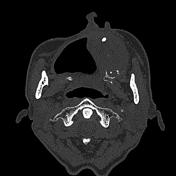
- peripheral nerve sheath tumour of mandible
- traumatic bone cyst of jaw (simple cyst)
- Stafne cyst
- brown tumour
- Langerhans cell histiocytosis
- Gorham disease
See also
-<li><a href="/articles/keratocystic-odontic-tumour">odontogenic keratocyst (keratocystic odontic tumour)</a></li>- +<li><a href="/articles/odontogenic-keratocyst">odontogenic keratocyst (keratocystic odontic tumour)</a></li>
- +<li><a title="Langerhans cell histiocytosis (skeletal manifestations)" href="/articles/langerhans-cell-histiocytosis-skeletal-manifestations-1">Langerhans cell histiocytosis</a></li>
- +<li><a title="Gorham disease" href="/articles/gorham-disease">Gorham disease</a></li>
References changed:
- 5. Dy AES, Cabungcal ACA, Pangan RM. Langerhans cell histiocytosis: vanishing mandible in a 10-year-old. (2016) Acta Oto-Laryngologica Case Reports. 1 (1): 36-39. <a href="https://doi.org/10.1080/23772484.2016.1209639">doi:10.1080/23772484.2016.1209639</a>
- 6. Raghuveer HP, Jayalekshmy R. Gorham's massive osteolysis of the mandible - a progressive radiographic presentation. (2009) Dento maxillo facial radiology. 38 (5): 292-5. <a href="https://doi.org/10.1259/dmfr/73198793">doi:10.1259/dmfr/73198793</a> - <a href="https://www.ncbi.nlm.nih.gov/pubmed/19474257">Pubmed</a> <span class="ref_v4"></span>
Images Changes:
Image 5 CT (bone window) ( update )
Caption
was changed:
Case 4: peripheral nerve sheath tumourtumor
Image 6 CT (bone window) ( update )

Caption
was changed:
Case 5: keratocystic odontic tumourtumor


 Unable to process the form. Check for errors and try again.