
Cases
By sharing our collective experience through interesting and classic patient cases, we can make a real difference in how people are imaged and diagnosed. Each case belongs to a contributing member and all cases are reviewed by our dedicated editors to ensure they reach quality standards and abide by privacy guidelines. Cases can public or unlisted and then be viewed directly or added to articles, playlists or multiple choice questions. Find out more about cases.


68 results found
Case
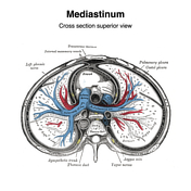
Mediastinum (Gray's illustrations)
Published
27 Feb 2024
35% complete
Diagram
Case
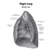
Pulmonary hila (Gray's illustrations)
Published
27 Feb 2024
32% complete
Diagram
Case
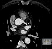
Retroartic left main coronary artery

Published
31 Jul 2023
87% complete
CT
Diagram
Case
Right coronary artery arising from the pulmonary artery

Published
31 Jul 2023
92% complete
DSA (angiography)
CT
Diagram
Case
Prepulmonic right coronary artery

Published
30 Jul 2023
87% complete
CT
Diagram
Case
Right coronary artery to right atrial fistula

Published
29 Jul 2023
83% complete
Diagram
CT
Case
Spotty coronary artery calcifications

Published
20 Jul 2023
77% complete
Diagram
CT
Case
Retroaortic circumflex coronary artery course

Published
20 Jul 2023
79% complete
Diagram
Case
Positive remodeling and napkin ring sign involving the right coronary artery

Published
20 Jul 2023
92% complete
DSA (angiography)
Diagram
CT
Case
"Malignant" anomalous interarterial course of the right coronary artery
Published
19 Jul 2023
87% complete
Diagram
CT
Case
Single coronary artery with prepulmonic left main coronary

Published
17 Jul 2023
84% complete
CT
Diagram
Case
Myocardial performance index formula

Published
15 Jun 2023
29% complete
Diagram
Case
Pericardial recesses

Published
10 Apr 2023
29% complete
Diagram
Case
Cardiac conduction device lead dislodgement (illustrations)

Published
24 Jan 2023
32% complete
Diagram
Case
Pacer pads and automated implantable cardioverter defibrillator in intubated patient

Published
22 Sep 2022
88% complete
Diagram
X-ray
Case
Left main coronary artery arising from the right pulmonary artery

Published
15 Mar 2022
87% complete
X-ray
CT
MRI
Diagram
Case
COVID-19 pneumonia and biosensor device

Published
14 Oct 2021
94% complete
X-ray
Photo
Diagram
Case
Truncal venous development (Gray's illustrations)

Published
15 Sep 2021
35% complete
Diagram
Case
Sinus venosus development (Gray's illustration)

Published
15 Sep 2021
29% complete
Diagram
Case
Aortic arches (Gray's illustration)

Published
14 Sep 2021
32% complete
Diagram
ADVERTISEMENT: Supporters see fewer/no ads


 Unable to process the form. Check for errors and try again.