


3,674 results found
Case
Basilar artery branches (illustration)

Published
15 May 2015
29% complete
Diagram
Case
Vidian artery

Published
15 May 2015
24% complete
CT
Case
Vertebrobasilar dolichoectasia (VDBE)

Published
15 May 2015
18% complete
CT
Case
Brain tumor frequency (chart)

Published
15 May 2015
29% complete
Diagram
Case
Salvador Dali (photo)

Published
15 May 2015
25% complete
Photo
Case
Hydatid cyst (photo)

Published
15 May 2015
29% complete
Photo
Case
Hydatid disease

Published
15 May 2015
19% complete
Photo
Case
Brain tumor incidence (graph)

Published
15 May 2015
19% complete
Diagram
Case
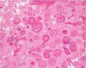
Echinococcus granulosus - scolex (photo)

Published
15 May 2015
29% complete
Pathology
Case
Parieto-occipital region (diagram)

Published
15 May 2015
32% complete
Diagram
Case
Meyer loop field cut (illustration)

Published
15 May 2015
19% complete
Diagram
Case
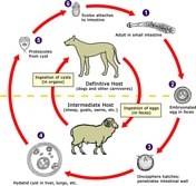
Echinococcus life cycle (diagram)
Published
15 May 2015
29% complete
Diagram
Case
Fetal posterior communicating artery

Published
15 May 2015
27% complete
CT
Case
Anterior cerebral artery terminal branches

Published
15 May 2015
27% complete
CT
Case
Bovine arch - illustration

Published
15 May 2015
33% complete
CT
Case
Anterior circle of Willis (illustration)

Published
15 May 2015
29% complete
Diagram
Case
Pericallosal moustache

Published
15 May 2015
29% complete
DSA (angiography)
Case
Lateral view of anterior cerebral artery

Published
15 May 2015
29% complete
DSA (angiography)
Case
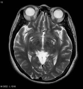
Perivascular spaces
Published
15 May 2015
53% complete
MRI
Case
Choroidal blush

Published
15 May 2015
25% complete
DSA (angiography)
Case
Vidian canal

Published
15 May 2015
33% complete
CT
Case
Betz cell of the primary motor cortex (histology)

Published
15 May 2015
29% complete
Pathology
Case
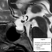
Supraoptic and infundibular recesses
Published
15 May 2015
22% complete
Annotated image
Case
Internal carotid artery segments (illustration)

Published
15 May 2015
19% complete
Annotated image
Case
Forceps minor

Published
15 May 2015
18% complete
MRI
Case
Forceps major

Published
15 May 2015
18% complete
MRI
Case
Anterior and middle cerebral arteries (annotated cerebral angiogram)

Published
15 May 2015
44% complete
Annotated image
Case
Cerebral vascular territories in the midline (illustration)

Published
15 May 2015
29% complete
Diagram
Case
Vascular territories of the lateral cerebral cortex (illustration)

Published
15 May 2015
32% complete
Diagram
Case
Eustachian tube (illustration)

Published
14 May 2015
29% complete
Diagram
Case
Eustachian tube on CT

Published
14 May 2015
24% complete
CT
Case
Eustachian tube (illustration)

Published
14 May 2015
32% complete
Diagram
Case
Distribution of paragangliomas (diagram)

Published
14 May 2015
29% complete
Diagram
Case
Haller cell and concha bullosa

Published
14 May 2015
56% complete
CT
Case
Facet joint injection - Scotty dog

Published
14 May 2015
19% complete
Fluoroscopy
Case
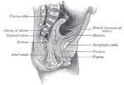
Female reproductive system
Published
14 May 2015
35% complete
Diagram
Case
Meningohypophyseal trunk

Published
14 May 2015
29% complete
DSA (angiography)
Case
Normal uterus

Published
14 May 2015
25% complete
Diagram
Case
Vascularization of the female reproductive system (Gray's illustration)

Published
14 May 2015
35% complete
Diagram
Case
Left temporal bone (illustration)

Published
14 May 2015
25% complete
Diagram
Case
Cerebral arterial supply to the brain (illustration)

Published
14 May 2015
25% complete
Diagram
Case
Carotid artery (illustration)

Published
14 May 2015
29% complete
Diagram
Case
ICA lateral post SAH vasospasm

Published
14 May 2015
35% complete
DSA (angiography)
Case
Vertebrobasilar angiography

Published
14 May 2015
32% complete
DSA (angiography)
Case
Fundus of internal acoustic meatus

Published
14 May 2015
25% complete
Diagram
Case
Angular gyrus and supramarginal gyrus (diagram)

Published
14 May 2015
22% complete
Diagram
Case
Exostosis of the ear canal

Published
14 May 2015
48% complete
CT
Case
Papillary thyroid carcinoma
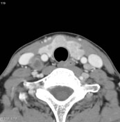
Published
14 May 2015
89% complete
CT
Case
Increased globe size (diagram)

Published
14 May 2015
29% complete
Diagram
Case
Evolution of MRI signal characteristics of intracranial hemorrhage (diagram)

Published
14 May 2015
19% complete
Diagram
Case
Evolution of CT density of intracranial hemorrhage (diagram)

Published
14 May 2015
32% complete
Diagram
Case
Globe (illustration)

Published
14 May 2015
44% complete
Diagram
Case
Broca's and Wernicke's areas (illustration)
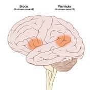
Published
14 May 2015
35% complete
Diagram
Case
Frontotemporal degeneration (gross pathology)
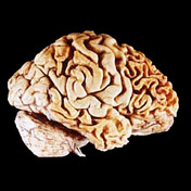
Published
14 May 2015
25% complete
Pathology
Case
Vertebral arteries (illustration)

Published
14 May 2015
27% complete
CT
Case
Adult polycystic kidney (gross pathology)

Published
14 May 2015
44% complete
Pathology
Case
Central neurocytoma (histology)

Published
14 May 2015
29% complete
Pathology
Case
Rosenthal fibers (histology)

Published
14 May 2015
29% complete
Pathology
Case
Apple core sculpture (photo)

Published
14 May 2015
25% complete
Photo
Case
Colorectal adenocarcinoma (gross pathology)

Published
14 May 2015
47% complete
Pathology
Case
Geniculate ganglion (illustration)
Published
13 May 2015
32% complete
Diagram
Case
Celery stalk (photo)

Published
13 May 2015
29% complete
Photo
Case
Sergeant stripes (diagram)

Published
13 May 2015
27% complete
Diagram
Case
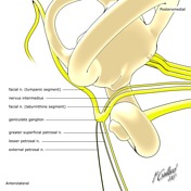
Nerves of the internal acoustic meatus (diagram)

Published
13 May 2015
32% complete
Diagram
Case
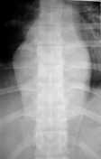
Pott disease
Published
13 May 2015
22% complete
X-ray
Case
Salivary gland tumor subtypes (table)

Published
13 May 2015
29% complete
Diagram
Case
Pinecone (photo)
Published
13 May 2015
25% complete
Photo
Case
Bilateral distal clavicular erosion

Published
13 May 2015
88% complete
X-ray
Case
Arnold's and Jacobson's nerves (diagram)

Published
13 May 2015
19% complete
Diagram
Case
Aortic intramural hematoma

Published
13 May 2015
77% complete
CT
Case
Linguine with tomato sauce (photo)

Published
13 May 2015
25% complete
Photo
Case
Pterygopalatine ganglion (illustration)

Published
13 May 2015
29% complete
Diagram
Case
Spinal arteriovenous malformation

Published
13 May 2015
74% complete
MRI
Case
Arteriovenous malformation - cerebral

Published
13 May 2015
92% complete
MRI
Case
Sacroiliitis - grade III

Published
13 May 2015
44% complete
X-ray
Case
Normal sacroiliac joint

Published
13 May 2015
19% complete
X-ray
Case
Target (photo)

Published
12 May 2015
25% complete
Photo
Case
Distal femoral avascular necrosis

Published
12 May 2015
59% complete
MRI
Case
Doughnut (photo)

Published
12 May 2015
29% complete
Photo
Case
Recurrent artery of Heubner infarct

Published
12 May 2015
59% complete
CT
Case
Funnel (photo)

Published
12 May 2015
32% complete
Photo
Case
Congenital pulmonary arteriovenous malformation

Published
12 May 2015
48% complete
CT
Case
Pineal tumor calcification (illustration)

Published
12 May 2015
25% complete
Diagram
Case
Pear (photo)

Published
12 May 2015
29% complete
Photo
Case
Bamboo (photo)

Published
12 May 2015
29% complete
Photo
Case
Sarcoidosis

Published
12 May 2015
69% complete
X-ray
Case
Banana (photo)

Published
12 May 2015
25% complete
Photo
Case
Medusa (photo)

Published
12 May 2015
25% complete
Photo
Case
Life cycle of Taenia solium (illustration)

Published
12 May 2015
32% complete
Diagram
Case
Palm tree (photo)

Published
12 May 2015
25% complete
Photo
Case
Blueberry (photo)

Published
12 May 2015
25% complete
Photo
Case
Nail-patella syndrome

Published
12 May 2015
47% complete
Annotated image
Case
Psammoma bodies - meningioma (histology)
Published
12 May 2015
32% complete
Pathology
Case
High grade astrocytoma (MRS)

Published
12 May 2015
65% complete
MRI
ADVERTISEMENT: Supporters see fewer/no ads


 Unable to process the form. Check for errors and try again.