


500 results found
Case
Normal orthopantomogram (OPG)

Published
16 Jan 2016
35% complete
X-ray
Case
Normal pelvic bone CT (age 16)

Published
06 Jul 2016
48% complete
CT
Case
Normal petrous temporal bone CT

Published
28 Nov 2014
53% complete
CT
Case
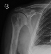
Normal shoulder MRI

Published
05 Mar 2016
45% complete
MRI
Case
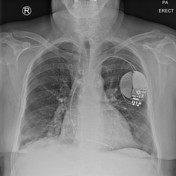
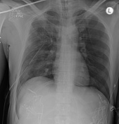
Normal supine trauma chest x-ray

Published
15 Oct 2014
54% complete
X-ray
Case
Normal supine trauma chest x-ray in an obese woman

Published
19 Oct 2014
44% complete
X-ray
Case
Normal supine trauma chest x-ray with rotation
Published
15 Oct 2014
85% complete
X-ray
Case
Normal thymus and pericardial recess

Published
24 May 2016
62% complete
CT
Case
Normal thymus on trauma CT

Published
24 May 2016
48% complete
CT
Case
Normal trauma cervical spine MRI
Published
16 Nov 2014
42% complete
MRI
Case
Normal trauma chest and pelvis x-rays

Published
15 Oct 2014
75% complete
X-ray
Case
Normal trauma chest, pelvis and spine imaging

Published
15 Oct 2014
81% complete
X-ray
CT
Case
Normal trauma series x-rays

Published
16 Oct 2014
47% complete
X-ray
Case
Normal trauma spine imaging

Published
25 Nov 2014
50% complete
CT
X-ray
Case
Normal trauma spine imaging (age 16)

Published
26 May 2016
45% complete
X-ray
CT
Case
Normal wrist CT including 3D

Published
22 May 2016
40% complete
CT
Case
Normal wrist MRI

Published
17 Feb 2016
42% complete
MRI
Case
Normal wrist x-rays

Published
04 May 2015
38% complete
X-ray
Case
Occipital condyle fracture

Published
25 Nov 2014
89% complete
CT
Case
Occipital extradural hematoma simulating venous sinus thrombosis

Published
06 Jul 2016
71% complete
CT
Case
Odontogenic keratocyst - pediatric

Published
03 Aug 2010
86% complete
X-ray
Case
Esophageal dilatation due to obstructing gastric band

Published
01 Jul 2011
75% complete
X-ray
Case
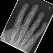
Old avulsion fracture at metacarpophalangeal joint

Published
01 May 2017
54% complete
X-ray
Case
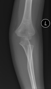
Olecranon foramen
Published
15 Feb 2019
79% complete
X-ray
Case
Omental torsion related to inguinal hernia

Published
07 Jun 2012
77% complete
CT
Case
Open book pelvic injury

Published
23 Oct 2014
85% complete
X-ray
Case
Os acromiale with impingement

Published
12 Apr 2010
87% complete
MRI
X-ray
Case
Os calcaneus secundarius

Published
02 Sep 2010
89% complete
CT
Case
Osteochondral fracture of the talar dome

Published
18 Jan 2015
88% complete
Annotated image
X-ray
Diagram
Case
Osteochondroma

Published
16 Aug 2013
100% complete
MRI
Case
Osteomyelitis

Published
23 Aug 2010
65% complete
X-ray
Nuclear medicine
MRI
Case
Osteoporosis circumscripta

Published
25 Aug 2018
83% complete
CT
Case
Ovarian fibroma (gross pathology)

Published
01 May 2010
41% complete
Pathology
Case
Ovarian mucinous cystadenoma (gross pathology)

Published
01 May 2010
41% complete
Photo
Case
Ovarian torsion (gross pathology)

Published
01 May 2010
32% complete
Photo
Case
Ovarian torsion with dermoid cyst
Published
28 Apr 2010
75% complete
Ultrasound
Case
Ovarian torsion with twisted pedicle

Published
01 May 2010
86% complete
Ultrasound
CT
Case
Paget disease of proximal humerus
Published
27 Feb 2020
82% complete
X-ray
Case
Paget disease: with blade of grass sign

Published
03 Aug 2014
91% complete
Annotated image
X-ray
CT
MRI
Nuclear medicine
Case
Pancreatic carcinoma with portal vein invasion

Published
16 Aug 2010
61% complete
Nuclear medicine
CT
Case
Parotid adenoidcystic carcinoma with carotid space spread

Published
19 Jul 2019
89% complete
MRI
Case
Parrot beak lateral meniscus tear and bucket handle medial mensicus tear

Published
26 Aug 2018
65% complete
MRI
Case
Partial anomalous pulmonary venous return (left upper lobe)
Published
25 Jul 2011
86% complete
CT
Case
Patchy right lower lobe consolidation
Published
27 Nov 2019
69% complete
X-ray
Case
Patella fracture simulating bipartite patella

Published
03 Oct 2016
92% complete
X-ray
CT
Case
Patellar instability risk factor assessment (diagram)

Published
15 Feb 2012
16% complete
Diagram
Case
Patellar tendinosis

Published
10 Apr 2010
92% complete
MRI
X-ray
Case
Patella tendon lateral femoral condyle friction syndrome

Published
08 Aug 2010
80% complete
MRI
Case
Pelvic bleeding treated with embolization

Published
27 Nov 2014
89% complete
CT
DSA (angiography)
Case
Pelvic fractures

Published
05 Nov 2014
89% complete
X-ray
CT
Case
Pelvic fractures (Malgaigne fracture)

Published
18 Oct 2014
92% complete
Annotated image
X-ray
CT
Case
Pelvic fracture with extraperitoneal hemorrhage

Published
24 May 2016
89% complete
CT
X-ray
Case
Penetrating wood foreign body injury to axilla

Published
27 May 2016
52% complete
CT
Case
Penetrating wood injury to axilla

Published
27 May 2016
86% complete
CT
Case
Peptic ulcer with early localized perforation

Published
04 May 2015
74% complete
CT
Case
Pericarditis

Published
19 Apr 2015
77% complete
CT
Case
Perifissural nodule

Published
05 Aug 2014
67% complete
CT
Annotated image
Case
Peroneus longus tendon rupture associated with an os peroneum

Published
26 Jul 2014
92% complete
Annotated image
X-ray
MRI
Case
Pes anserinus bursa (diagram)

Published
27 Feb 2012
25% complete
Annotated image
Case
Pes anserinus tendon (diagram)

Published
26 Feb 2012
25% complete
Annotated image
Diagram
Case
PHACE syndrome

Published
13 Aug 2010
68% complete
MRI
Case
Pharyngeal pouch

Published
18 Jul 2010
80% complete
X-ray
Barium
CT
Case
'Pill cam' related small bowel perforation

Published
21 Jun 2011
90% complete
X-ray
CT
Case
Pineal cyst

Published
18 Sep 2011
74% complete
CT
Case
Pituitary macroadenoma

Published
24 May 2012
56% complete
MRI
Case
Placental abruption

Published
12 Jul 2011
66% complete
Ultrasound
Case
Planum sphenoidale meningioma

Published
19 Jan 2015
65% complete
CT
Case
Pneumocystis pneumonia

Published
20 Jul 2011
54% complete
X-ray
Case
Pneumocystis pneumonia

Published
04 May 2010
97% complete
X-ray
Case
Pneumoperitoneum

Published
22 Apr 2015
91% complete
Annotated image
X-ray
Case
Pneumothorax and pelvic fractures

Published
15 Oct 2014
91% complete
X-ray
Case
Pneumothorax on expiratory radiograph

Published
03 Oct 2016
88% complete
X-ray
Case
Pneumothorax with deep sulcus sign
Published
08 Sep 2017
85% complete
X-ray
Case
Polyarticular foot gout on dual energy CT

Published
25 Aug 2018
77% complete
CT
X-ray
Case
Pontine infarction

Published
14 Jul 2011
77% complete
CT
MRI
Case
Popliteal artery entrapment syndrome

Published
15 Apr 2010
92% complete
MRI
Case
Popliteus tendon gout on MRI

Published
25 Aug 2018
74% complete
MRI
Case
Posterior cruciate ligament tear and associated injuries

Published
08 Jul 2015
92% complete
MRI
Case
Posterior dislocation of sternoclavicular joint

Published
27 Feb 2020
89% complete
CT
X-ray
Case
Posterior shoulder dislocation with reverse Hill-Sachs

Published
26 Feb 2020
86% complete
X-ray
Case
Posterior shoulder dislocation with reverse Hill-Sachs and reverse Bankart lesions

Published
29 Nov 2010
89% complete
X-ray
CT
Case
Posterior urethral valves with pyonephrosis

Published
13 Aug 2010
63% complete
Fluoroscopy
Ultrasound
Case
Prepatellar gout on dual energy CT

Published
25 Aug 2018
83% complete
X-ray
CT
Case
Prominent cervical vertebral venous channels

Published
24 May 2016
86% complete
CT
Case
Prominent pericardial recess on trauma CT

Published
11 Sep 2017
74% complete
CT
Case
Prominent perivascular space

Published
14 Apr 2016
87% complete
CT
MRI
Case
Proximal femoral fracture

Published
19 Oct 2014
88% complete
X-ray
Case
Proximal humeral metastasis

Published
27 Feb 2020
82% complete
X-ray
Case
Proximal phalanx osteomyelitis following laceration
Published
21 Oct 2019
82% complete
X-ray
Case
Proximal radial shaft fracture with radial head subluxation
Published
19 Apr 2015
91% complete
X-ray
Case
Proximal rupture of long head biceps brachii tendon

Published
14 Jan 2016
86% complete
MRI
Case
Pseudoarthrosis of the clavicle

Published
17 Aug 2010
65% complete
X-ray
CT
Case
Pseudo-subarachnoid hemorrhage

Published
07 Apr 2010
80% complete
CT
Case
Pubic ramus stress fracture

Published
09 Aug 2016
86% complete
MRI
ADVERTISEMENT: Supporters see fewer/no ads


 Unable to process the form. Check for errors and try again.